
Breast Cancer Treatment in Aurangabad — Expert Care at Karnawat Cancer Hospital
- MCh Surgical Oncologist — Cancer Surgery Specialist
- DM Medical Oncologist — Gold Medalist
- Breast-Conserving Surgery Available
- Onco-Plastic Reconstruction
- Aurangabad, Marathwada
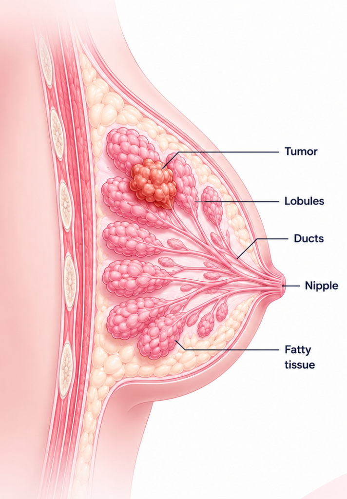
What Is Breast Cancer?
Breast cancer is the most common cancer in women in India — and it is highly treatable when detected early. Stage 1 breast cancer has a five-year survival rate greater than 95%. This is why awareness, regular screening, and early consultation with a breast cancer specialist are so important. At Karnawat Cancer Hospital in Aurangabad, breast cancer is treated with a multidisciplinary approach — surgery, chemotherapy, targeted therapy, hormone therapy, and onco-plastic reconstruction — all coordinated by specialists in one dedicated centre.
Breast Cancer Statistics in India & Maharashtra
1.7 Lakh+
New cases of breast cancer in India every year
14+
of all cancers in Indian women
95%+
5-year survival rate in early detection (Stage 1)
Marathwada
needs more specialised breast cancer care
Types of Breast Cancer
Based on Location of Origin
Invasive Ductal Carcinoma (IDC):
The most common type, accounting for about 70–80% of all breast cancers. Cancer cells start in the milk ducts and invade the surrounding breast tissue.
Invasive Lobular Carcinoma (ILC):
Begins in the lobules. About 10–15% of invasive breast cancers. Can be harder to detect on mammography.
Ductal Carcinoma In Situ (DCIS):
A non-invasive (Stage 0) cancer where abnormal cells are confined within milk ducts. Highly curable but requires treatment to prevent progression.
Lobular Carcinoma In Situ (LCIS):
Not a true cancer but a marker of increased risk. Requires monitoring.
Based on Molecular Subtype (Receptor Status)
Hormone Receptor-Positive (HR+):
Cancer cells have receptors for estrogen (ER+) or progesterone (PR+). Respond to hormone-blocking therapies like tamoxifen or aromatase inhibitors. Most common subtype.
HER2-Positive (HER2+):
Cancer overproduces HER2 protein, driving aggressive growth. Treated with targeted drugs such as trastuzumab (Herceptin) and pertuzumab (Perjeta).
Triple-Negative Breast Cancer (TNBC):
Tests negative for ER, PR, and HER2. Most aggressive subtype. Treated with chemotherapy, immunotherapy (pembrolizumab), and targeted agents like olaparib for BRCA-mutated TNBC.
Luminal A:
ER+/PR+, HER2-negative, low Ki-67 (slow-growing). Best prognosis. Often treated with hormone therapy alone.
Luminal B:
ER+, HER2+ or high Ki-67 (faster-growing). Requires more aggressive treatment including chemotherapy.
Less Common Breast Cancer Types
Inflammatory Breast Cancer (IBC):
Rare but aggressive. Does not cause a typical lump — breast appears red, swollen, warm. Requires urgent specialist evaluation.
Paget’s Disease of the Breast:
Rare cancer affecting the skin of the nipple and areola.
Symptoms
- A new lump or thickening in the breast or underarm that does not go away — even if painless
- Change in the size, shape, or contour of the breast
- Skin changes: dimpling, puckering, redness, or orange-peel texture (peau d’orange)
- Nipple inversion — a nipple that suddenly turns inward
- Nipple discharge — especially if bloody or occurring without squeezing
- Scaling, peeling, crusting, or flaking of the nipple skin
- Persistent breast pain or tenderness that is new and unexplained
- Swelling of part of the breast, even without a distinct lump
- Redness or warmth of the breast skin (may indicate inflammatory breast cancer)
- Visible veins on the breast surface that were not there before
Risk Factors
Risk Factors You Cannot Change
- Being female: Women are 100 times more likely than men to develop breast cancer
- Age: Risk increases with age; most cases are diagnosed after 50
- Family history: First-degree relative with breast cancer roughly doubles risk
- BRCA1 / BRCA2 gene mutations: Increase lifetime risk up to 70%; genetic testing available at Karnawat Cancer Hospital
- Personal history of breast cancer increases risk in the other breast
- Dense breast tissue on mammography
- Previous radiation therapy to the chest
- Early menstruation (before age 12) or late menopause (after 55)
Modifiable Risk Factors
- Alcohol consumption — risk rises with amount consumed
- Obesity after menopause — excess fat increases estrogen levels
- Physical inactivity
- Combined estrogen-progesterone HRT for more than 5 years
- Late first pregnancy or never having children
- Not breastfeeding — breastfeeding has a protective effect
Screening
Breast Self-Examination (BSE)
Screening by Age
| Age Group | Recommendation |
|---|---|
| 20–39 Years | Clinical breast exam every 1–3 years; monthly Breast Self-Examination (BSE); BRCA testing if there is a family history. |
| 40–44 Years | Discuss mammography with your doctor based on your personal risk factors. |
| 45–54 Years | Annual mammography is recommended. |
| 55+ Years | Annual or biennial mammography depending on your overall risk profile. |
| High-Risk (BRCA+) | Annual mammography with Breast MRI starting around age 30 or as advised by your oncologist. |
Diagnosis
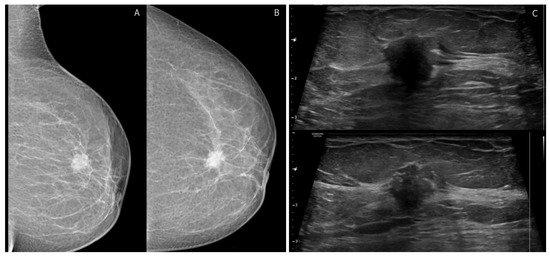
Step 1 — Imaging
Mammography: X-ray identifying suspicious masses, calcifications, or distortions.
Breast Ultrasound: Differentiates solid masses from cysts. Useful for younger women with denser breast tissue.
MRI: Detailed imaging for staging, extent of disease, and high-risk screening.
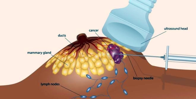
Step 2 — Biopsy
FNAC: Fine needle aspiration of cells. Quick but limited information.
Core Needle Biopsy (CNB): Standard at Karnawat Hospital. Provides full pathological assessment including hormone receptor and HER2 testing.
Excision Biopsy: Surgical removal of lump when needle biopsy is inconclusive.
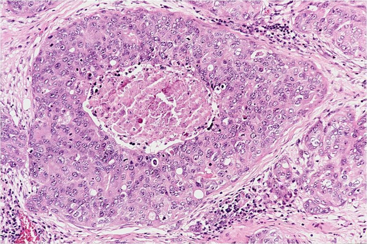
Step 3 — Pathology Report
Reports: cancer type, grade, hormone receptor status (ER, PR), HER2 status, and Ki-67 (cell proliferation rate). This directly determines your treatment plan.
Step 4 — Staging Investigations
Blood tests, CT scan chest/abdomen/pelvis, bone scan or PET-CT, and brain MRI in select cases.
Staging
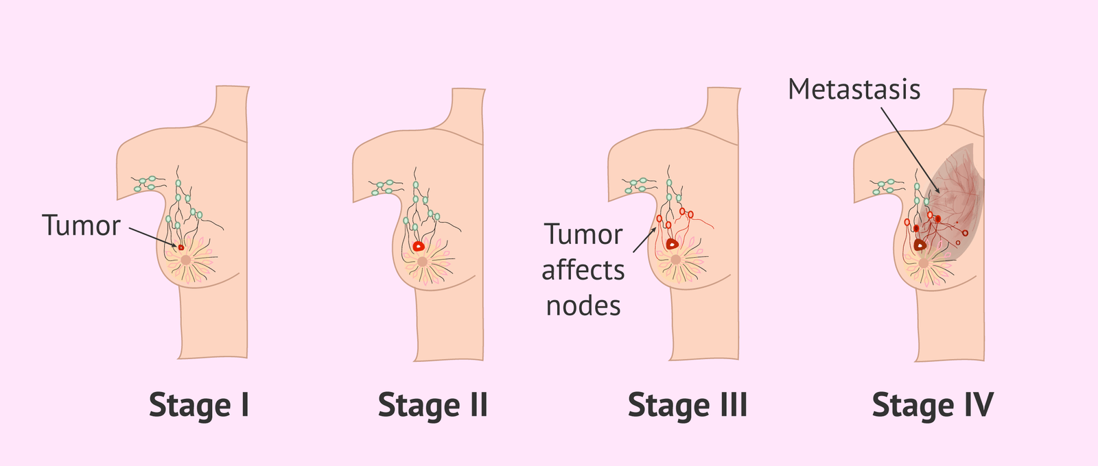
| Stage | Description | 5-Year Survival (Approx.) |
|---|---|---|
| Stage 0 (DCIS) | Cancer cells confined within milk ducts, not invasive. | ~99% |
| Stage 1 | Small tumour (≤2 cm), no or minimal lymph node involvement. | ~95–99% |
| Stage 2 | Tumour 2–5 cm and/or spread to 1–3 axillary lymph nodes. | ~75–90% |
| Stage 3 | Tumour larger than 5 cm, spread to four or more lymph nodes, or involvement of the skin/chest wall. | ~50–70% |
| Stage 4 (Metastatic) | Cancer has spread to distant organs such as the lungs, liver, bones, or brain. | ~25–30% (Improving with modern therapies) |
Stage 0 (DCIS)
Cancer cells confined within milk ducts, not invasive.
Stage 1
Small tumour (≤2 cm), no or minimal lymph node involvement.
Stage 2
Tumour 2–5 cm and/or spread to 1–3 axillary lymph nodes.
Stage 3
Tumour larger than 5 cm, spread to four or more lymph nodes, or involvement of the skin/chest wall.
Stage 4 (Metastatic)
Cancer has spread to distant organs such as the lungs, liver, bones, or brain.
Treatment Options Overview
- Surgery — removes the tumour
- Radiation therapy — destroys residual cancer cells locally
- Chemotherapy — kills cancer cells systemically
- Targeted therapy — attacks specific cancer cell molecules
- Hormone therapy — blocks hormones that fuel cancer growth
- Immunotherapy — activates immune system against cancer
- Breast reconstruction — restores appearance after mastectomy
Surgical Treatment for Breast Cancer
Breast-Conserving Surgery (Lumpectomy)
Removes the tumour and a margin of surrounding normal tissue while leaving the rest of the breast intact. Suitable for Stage 1 and Stage 2 cancers where tumour is small relative to breast size. Almost always followed by radiation therapy.
Mastectomy
Removes the entire breast tissue. Recommended for large tumours, multiple tumours, recurrence, BRCA mutation carriers, or patient preference.
Sentinel Lymph Node Biopsy (SLNB)
Removes only the first lymph node(s) to which breast cancer would spread. If cancer-free, full axillary dissection is avoided — significantly reducing lymphedema risk. Standard of care at Karnawat Cancer Hospital.
Neoadjuvant Approach (Chemotherapy Before Surgery)
For locally advanced tumours or HER2-positive/triple-negative cancers, chemotherapy may be given before surgery to shrink the tumour, potentially making breast-conserving surgery possible and assessing treatment response. The decision is made jointly by both specialists — a key benefit of Karnawat Hospital’s dual-specialist structure.
Onco-Plastic Breast Surgery — Karnawat’s Unique Advantage
This is a key differentiator for Karnawat Cancer Hospital in Marathwada. Onco-plastic surgery combines cancer removal with plastic surgery techniques to achieve the best possible cosmetic outcome — complete cancer excision AND aesthetic restoration in one procedure.
Standard lumpectomy can leave visible deformity when removing a large tumour. Onco-plastic techniques reshape the remaining breast tissue or balance with a contralateral breast reduction, preserving body confidence and self-image. Dr. Anand Karnawat’s fellowship training in Microvascular Surgery makes this available in Marathwada.
| Mastectomy Type | What Is Removed | Reconstruction Option |
|---|---|---|
| Simple (Total) Mastectomy | All breast tissue including nipple-areola; no lymph nodes | Yes |
| Modified Radical Mastectomy | All breast tissue + axillary lymph nodes | Yes |
| Skin-Sparing Mastectomy | Breast tissue; breast skin preserved | Yes — excellent result |
| Nipple-Sparing Mastectomy | Breast tissue only; nipple-areola preserved | Yes — best cosmetic result |
Medical Oncology Treatment
Chemotherapy for Breast Cancer
May be used before surgery (neoadjuvant), after surgery (adjuvant), or for metastatic disease. Common regimens include:
- AC (Adriamycin + Cyclophosphamide) — standard anthracycline-based regimen
- Taxane-based regimens: Paclitaxel, Docetaxel — particularly effective in TNBC and HER2+ cancers
- AC-T Dose-Dense — intensive schedule for high-risk early breast cancer
- Carboplatin — added for TNBC or BRCA-mutated breast cancer
Immunotherapy
Most relevant for Triple-Negative Breast Cancer (TNBC) — PD-L1 positive:
- Pembrolizumab (Keytruda): Immune checkpoint inhibitor approved for PD-L1+ metastatic TNBC (with chemo) and high-risk early TNBC (neoadjuvant + adjuvant)
- Atezolizumab (Tecentriq): Another checkpoint inhibitor used in TNBC
Immunotherapy eligibility is determined by molecular testing including PD-L1 expression and tumour mutational burden (TMB).
Radiation Therapy
High-energy beams used after breast-conserving surgery (always), after mastectomy if tumour was large or nodes involved, and for metastatic disease symptom control. Where radiation is required, Karnawat Cancer Hospital coordinates with a trusted radiation oncology centre, with full continuity of treatment planning.
Targeted Therapy
HER2-Targeted Agents:
| Drug | Type | Used For |
|---|---|---|
| Trastuzumab (Herceptin) | HER2-targeted antibody | HER2+ early & metastatic BC; 12 months adjuvant |
| Pertuzumab (Perjeta) | HER2-targeted antibody | HER2+ high-risk early BC or metastatic |
| T-DM1 (Kadcyla) | Antibody-drug conjugate | Residual disease after neoadjuvant therapy |
| Lapatinib / Tucatinib | Oral HER2 inhibitors | Later-line metastatic HER2+ BC |
CDK4/6 Inhibitors (HR+/HER2- breast cancer):
- Palbociclib (Ibrance), Ribociclib (Kisqali), Abemaciclib (Verzenio) — combined with hormone therapy for metastatic HR+/HER2- BC and high-risk early BC.
PARP Inhibitors (BRCA-mutated breast cancer):
- Olaparib (Lynparza), Talazoparib (Talzenna) — for germline BRCA1/2-mutated HER2- metastatic BC; also adjuvant in high-risk early disease.
Hormone Therapy (Endocrine Therapy)
Used for ER+ and/or PR+ breast cancers — the most common subtype. Works by blocking estrogen from reaching cancer cells or reducing estrogen levels.
| Drug | Type | Suitable For |
|---|---|---|
| Tamoxifen | Estrogen receptor blocker | Premenopausal; 5–10 years |
| Letrozole / Anastrozole / Exemestane | Aromatase inhibitors | Postmenopausal; 5–10 years |
| Ovarian Suppression | Reduces ovarian estrogen | Premenopausal high-risk patients |
| Fulvestrant (Faslodex) | Estrogen receptor degrader | Metastatic HR+ breast cancer |
Breast Reconstruction After Cancer Surgery
Timing of Reconstruction
Immediate Reconstruction:
Performed at the same time as mastectomy. Patient wakes with a reconstructed breast mound.
Delayed Reconstruction:
Performed weeks to years after mastectomy. May be recommended if post-mastectomy radiation is planned.
Types of Reconstruction
| Type | Method | Key Benefit |
|---|---|---|
| Implant-Based | Tissue expander then silicone/saline implant | Simpler surgery, faster recovery, no donor site |
| TRAM Flap | Abdominal muscle and skin | Natural result, long-lasting |
| DIEP Flap | Abdominal skin and fat, NO muscle | Preserves abdominal strength; requires microvascular expertise |
| Latissimus Dorsi Flap | Upper back tissue | Sometimes combined with implant |
Dr. Anand Vinay Karnawat’s Fellowship in Microvascular Surgery means complex reconstruction options including DIEP flap are available at Karnawat Cancer Hospital — a rare capability in Marathwada.
Before Treatment: What to Prepare
Before Surgery
- Blood tests, ECG, and anaesthesia evaluation will be scheduled
- Blood thinners (aspirin, NSAIDs) stopped 7–10 days before surgery
- Fast from midnight before the procedure
- Arrange assistance at home for the first few days after surgery
- Bring a list of all medications and allergies to your pre-surgical appointment
Before Chemotherapy
- Dental check-up recommended
- Blood counts checked before each cycle
- Port placement may be recommended for intravenous chemotherapy access
- Arrange support at home on treatment days and the 5–7 days following
- Discuss anti-nausea medications and dietary guidance with your oncology team
Fertility Preservation (Premenopausal Women)
After Treatment — Recovery & Follow-Up
Post-Surgery Recovery
• Light activities resume within 2–4 weeks
• Drain tubes removed within 1–2 weeks
• Arm exercises important after axillary surgery — guided by our team
• Wound care instructions provided before discharge
Lymphedema
Swelling of the arm on the operated side can occur after axillary lymph node surgery. Early management with physiotherapy, compression garments, and lymphatic drainage massage is effective. Report any arm swelling to your oncologist promptly.
Follow-Up Schedule
Signs of Recurrence to Report Immediately
- New lump in treated breast, opposite breast, or axilla
- Persistent bone pain — especially back, hips, or ribs
- Persistent cough or shortness of breath
- Unexplained headaches or vision changes
- Unexplained weight loss or abdominal pain
After Treatment — Recovery & Follow-Up
| ❌ Myth | ✔ Fact |
|---|---|
| Only women with a family history get breast cancer | About 85% of breast cancers occur in women with no family history. All women face some risk. |
| A breast lump is always cancer | Most lumps (80–85%) are benign — fibroadenomas, cysts, or fibrocystic changes. Only a biopsy confirms cancer. |
| Mastectomy is always necessary | Lumpectomy + radiation gives equivalent survival to mastectomy for early-stage breast cancer. |
| Young women don’t get breast cancer | Breast cancer can occur in women in their 20s, 30s, and 40s. Age is a risk factor, not a guarantee. |
| Chemotherapy always causes hair loss | Not all regimens cause hair loss. It depends on specific drugs used. Hair regrows after treatment. |
| Mammography radiation causes cancer | The dose is extremely low. The benefit of early detection far outweighs this minimal risk. |
| After mastectomy, breast cancer cannot return | Recurrence can happen in the chest wall, lymph nodes, or distant organs. Regular follow-up is essential. |
| Men cannot get breast cancer | Men can develop breast cancer, though rare (~1% of all cases). Men with BRCA2 mutations are at increased risk. |
| Underwire bras / deodorants cause breast cancer | No scientific evidence links these to breast cancer. |
| Surgery makes cancer spread | Breast cancer surgery does not cause cancer to spread when performed by a trained surgical oncologist. |
Why Choose Karnawat Cancer Hospital?
The Only Dual-Specialist Cancer Centre in Marathwada
Karnawat Cancer Hospital is the only dedicated cancer centre in Marathwada where an MCh Surgical Oncologist and a DM Medical Oncologist are both permanently on-site. For breast cancer — which requires both surgical and medical oncology expertise — this integrated structure provides a genuine clinical advantage.
Onco-Plastic Surgery — Cancer Removal Without Sacrificing Appearance
Dr. Anand Karnawat’s training in microvascular and onco-plastic surgery means patients can have cancer removed with optimal cosmetic outcomes. For many women, this is a life-changing difference not widely available in Marathwada.
ESMO-Certified Medical Oncology
Dr. Khushboo Jain Karnawat holds ESMO (European Society for Medical Oncology) certification — signifying advanced oncology training and knowledge of international treatment guidelines. Treatment at Karnawat Hospital aligns with globally recognised standards.
Personalised Treatment Plans
Every breast cancer patient receives an individualised plan. Your cancer’s receptor status, grade, stage, fertility wishes, and personal circumstances are all considered. You are not assigned to a protocol. You are understood as a person.
Affordable Access to World-Class Oncology
Karnawat Cancer Hospital brings expert oncology to Aurangabad, eliminating the financial and physical burden of traveling to Pune or Mumbai for specialist cancer care.
Government Health Scheme Guidance
Our team guides patients on accessing Mahatma Phule Jan Arogya Yojana (MPJAY) and Ayushman Bharat (PMJAY) for eligible cancer treatments.
Meet the Breast Cancer Specialists

Dr. Anand Vinay Karnawat — Breast Cancer Surgeon in Aurangabad
MBBS | MS (General Surgery) | MCh (Surgical Oncology) | FMAS | FICRS | Fellowship in Microvascular Surgery Dr. Anand Karnawat is a formally trained Surgical Oncologist with extensive experience in breast cancer surgery — including lumpectomy, mastectomy, sentinel lymph node biopsy, axillary dissection, and onco-plastic reconstruction. His microvascular fellowship enables complex breast reconstruction procedures not widely available in Marathwada.
Book an Appointment:
+91 79721 85084
WhatsApp:
+91 99437 44480
OPD Hours:
Monday – Saturday | 9 AM – 6 PM
Address:
K Sector, Mayur Nagar, N-11, CIDCO, Chhatrapati Sambhajinagar (Aurangabad)

Dr. Khushboo Jain Karnawat — Breast Cancer Medical Oncologist in Aurangabad
MBBS | MD (Radiation Oncology) | DM (Medical Oncology) — Gold Medalist | DrNB | ECMO (ESMO Certified) Dr. Khushboo Jain Karnawat manages chemotherapy, HER2-targeted therapy (trastuzumab, pertuzumab), hormone therapy, CDK4/6 inhibitors, immunotherapy, and all systemic drug treatments for breast cancer. As an ESMO-certified oncologist, she applies internationally recognised guidelines to every patient’s care.
Frequently Asked Questions
Yes, especially when detected early. Stage 1 breast cancer has a five-year survival rate above 95%. Even Stage 2 and 3 breast cancers have excellent outcomes with appropriate treatment. Stage 4 is generally not curable but is increasingly manageable as a chronic disease, with many patients living 5–10+ years.
Clinical studies show lumpectomy + radiation therapy provides equivalent long-term survival to mastectomy for early-stage breast cancer. The choice depends on tumour size, location, breast size, BRCA status, and personal preference. Your surgeon will discuss options honestly for your specific case. Both are valid choices.
Hair loss depends on the specific chemotherapy drugs used. Anthracycline and taxane regimens (common in breast cancer) typically cause temporary hair loss — hair regrows within 3–6 months after treatment ends. Discuss which drugs are in your regimen and what to expect with your Medical Oncologist.
For hormone receptor-negative breast cancers, pregnancy after treatment is generally considered safe. For hormone receptor-positive cancers, most guidelines recommend completing hormone therapy (5–10 years) before attempting pregnancy — though guidance is evolving. Discuss fertility and family planning with your oncologist before treatment begins.
BRCA1 and BRCA2 mutations significantly increase lifetime breast cancer risk (up to 70%) and ovarian cancer risk. Testing is recommended if you have breast cancer at a young age, bilateral breast cancer, ovarian cancer in the family, or male relatives with breast cancer. Genetic counseling and testing are available at Karnawat Cancer Hospital.
How do I book an appointment?
Book an Appointment:
+91 79721 85084
WhatsApp:
+91 99437 44480
OPD Hours:
Monday – Saturday | 9 AM – 6 PM
Address:
K Sector, Mayur Nagar, N-11, CIDCO, Chhatrapati Sambhajinagar (Aurangabad)